Home
/ Deigram Of Outside Leg Muscles, Anatomy Of The Quadriceps Muscles : It contains the peroneus longus and peroneus brevis muscles.
Deigram Of Outside Leg Muscles, Anatomy Of The Quadriceps Muscles : It contains the peroneus longus and peroneus brevis muscles.
Deigram Of Outside Leg Muscles, Anatomy Of The Quadriceps Muscles : It contains the peroneus longus and peroneus brevis muscles.. The nerve signals in these reflexes come from stretch receptors located in the joints, ligaments, tendons, and even the muscles themselves. Muscle anatomy for gym 12 photos of the muscle anatomy for gym muscle anatomy and fitness, muscle anatomy for fitness, muscle anatomy for gym, human muscles, muscle anatomy and fitness, muscle anatomy for fitness, muscle anatomy for gym Extend the arm that is on the same side as your outstretched leg, and place it so the elbow presses against the outside of the bent knee. It contains the peroneus longus and peroneus brevis muscles. The muscles in the hip are responsible for the movement of the hip and, by proxy, the leg.
Extend the arm that is on the same side as your outstretched leg, and place it so the elbow presses against the outside of the bent knee. The anterior compartment of the leg acts to dorsiflex and invert the foot through the ankle joint. Human muscle system, the muscles of the human body that work the skeletal system, that are under voluntary control, and that are concerned with movement. The hamstring muscle attachment points. Observe the leg muscle diagram posted above and notice that there are many parts in the muscles.the largest muscle masses in the leg are present in the thigh and the calf.
Tight Calf Muscles Causes And Stretches For Tight Calf Muscles from www.sportsinjuryclinic.net Bring museum quality art into your home or office decor with a canvas print that will never warp or sag. However, many reflex pathways are also active in the legs and foot. The extensor hallucis longus and the extensor digitorum … Your quadricep muscles, also known as quads, consist of four muscles that compose the front of your leg; The top leg/foot should cross over the extended bottom leg near the knee. Included are several layered views of the back muscles, the dorsal muscles, subclavius muscles, rhomboideus major and minor muscles, deltoid muscles and many more. Extension, flexion, adduction, and abduction. The legs are the lower limbs of the human body that provide support and stability in addition to allowing movement.
Bring one leg over the opposite thigh, and place the foot on the floor.
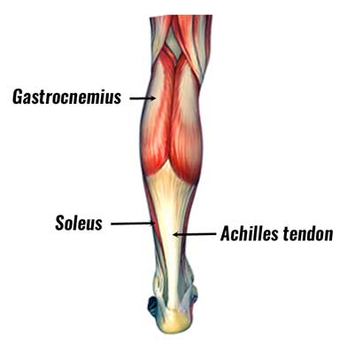
The muscles in the hip are responsible for the movement of the hip and, by proxy, the leg. Included are several layered views of the back muscles, the dorsal muscles, subclavius muscles, rhomboideus major and minor muscles, deltoid muscles and many more. Tibialis anterior, extensor digitorum longus, extensor hallicus longus, fibularis (peroneus) longus, fibu. It gets its blood flow from the arteries in the tiberial artery. To feel these muscles contract, place your hand on the outside of your shin and turn your foot out. The gastrocnemius muscle has two large bellies, called the medial head and the lateral head, and inserts into the calcaneus bone of the. This is why you have to indicate which biceps you are taking about when discussing one or other of these muscles. Biceps femoris (long head) biceps femoris (short head) semitendinosus. Supporting, balancing, and propelling the body is the work of the muscular system of the legs and feet. This muscle runs along the outside of the back of your thigh and attaches to the top of the fibula (the smaller of the two bones of your lower leg). Both comments and trackbacks are currently closed. The extensor hallucis longus and the extensor digitorum … The top leg/foot should cross over the extended bottom leg near the knee.
Notice the upper leg has a biceps muscle just like the upper arm does. The anterior compartment of the leg acts to dorsiflex and invert the foot through the ankle joint. Posterior compartment, also known as the flexor compartment; They also help with pointing the foot, or plantarflexion. The nerve signals in these reflexes come from stretch receptors located in the joints, ligaments, tendons, and even the muscles themselves.
Leg Anatomy And Function Of Bones And Muscles Plus Diagram from post.healthline.com The top leg/foot should cross over the extended bottom leg near the knee. The legs include the upper leg, knee, lower leg, ankle, and. The thigh (proximal lower limb) muscles are arranged into three compartments : Bring museum quality art into your home or office decor with a canvas print that will never warp or sag. Muscle and bone anatomy 12 photos of the muscle and bone anatomy back muscles and bones anatomy, human muscle and bone anatomy, muscle & bone anatomy 3d free download, muscle and bone anatomy app, muscle and bone anatomy quiz, human muscles, back muscles and bones anatomy, human muscle and bone anatomy, muscle & bone. Muscles of the leg and foot. These are the muscles that are located there: The muscles that make up the quadriceps are the strongest and leanest of all muscles in the body.
This is the biggest muscle that is in the tibialis anterior.
The upper leg, in particular, is comprised of bones and muscles that are susceptible to injury, particularly when excess strain is placed upon them. On the outside of the thigh, this is the largest of. In the leg, muscle strains happen when a muscle is either stretched beyond its limits or forced into extreme contraction. The anterior compartment of the leg acts to dorsiflex and invert the foot through the ankle joint. Human muscle system, the muscles of the human body that work the skeletal system, that are under voluntary control, and that are concerned with movement. Add a frame to any. The calf muscle, on the back of the lower leg, is actually made up of two muscles: Reflexes help to maintain proper muscle tone, balance, and responsiveness of the legs and feet to stimuli such as stepping on a sharp object. This is the biggest muscle that is in the tibialis anterior. These muscles pull the toes and feet outward. This is why you have to indicate which biceps you are taking about when discussing one or other of these muscles. From the large, strong muscles of the buttocks and legs to the tiny, fine muscles of the feet and toes, these muscles can exert tremendous power while constantly making small adjustments for balance — whether. Your quadricep muscles, also known as quads, consist of four muscles that compose the front of your leg;
Anterior compartment leg muscles the anterior compartment of the leg comprises four muscles. Some of the more common ones are: Your quadricep muscles, also known as quads, consist of four muscles that compose the front of your leg; The muscles in the hip are responsible for the movement of the hip and, by proxy, the leg. Human muscle system, the muscles of the human body that work the skeletal system, that are under voluntary control, and that are concerned with movement.
Ohiodance Knee Anatomy from ohiodance.org The muscles in the upper leg power many of our movements. Bring museum quality art into your home or office decor with a canvas print that will never warp or sag. Related posts of lower leg muscles diagram muscle and bone anatomy. The thigh (proximal lower limb) muscles are arranged into three compartments : They also help with pointing the foot, or plantarflexion. Posterior compartment, also known as the flexor compartment; Extend the arm that is on the same side as your outstretched leg, and place it so the elbow presses against the outside of the bent knee. Some of the more common ones are:
This is the biggest muscle that is in the tibialis anterior.
Extend the arm that is on the same side as your outstretched leg, and place it so the elbow presses against the outside of the bent knee. These muscles pull the toes and feet outward. The muscles work together to enable movement and keep the hip in alignment. The hamstring muscles, also known as the rear thighs, make up the backside of the upper leg anatomy. Extension, flexion, adduction, and abduction. On the medial edge of the posterior thigh is the gracilis muscle. Included are several layered views of the back muscles, the dorsal muscles, subclavius muscles, rhomboideus major and minor muscles, deltoid muscles and many more. Muscle and bone anatomy 12 photos of the muscle and bone anatomy back muscles and bones anatomy, human muscle and bone anatomy, muscle & bone anatomy 3d free download, muscle and bone anatomy app, muscle and bone anatomy quiz, human muscles, back muscles and bones anatomy, human muscle and bone anatomy, muscle & bone. This muscle runs along the outside of the back of your thigh and attaches to the top of the fibula (the smaller of the two bones of your lower leg). Legs give us the freedom to run, walk, jump, climb, and negotiate the world around us. On the outside of the thigh, this is the largest of. This video identifies all muscles of the lower leg. This is the group of muscles that you often see body builders flexing, which protrude just above the knee and take up most of the upper leg.